Showing posts with label Ventricle. Show all posts

Monday, September 25, 2017

What is slit ventricle syndrome (SVS)?


I. Understanding our ventricular system
brain parenchyma

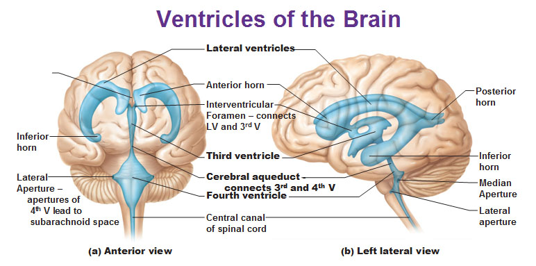
One error that we make in considering the ventricular system of the human brain is that it is static and that the ventricles do not modify over time. This is not true and evidence of this can be seen even before birth by viewing a prenatal ultrasound where they are relatively small. What is the ventricular system? Simply put, it is a communicating network of cavities filled with cerebrospinal fluid (CSF) located within the brain parenchyma (pictured at right). It is made up of two (2) lateral ventricles, the third ventricle (the site used for an endoscopic third ventriculostomy or ETV), the cerebral aqueduct, and the fourth ventricle. Additionally, the choroid plexuses - responsible for the production of CSF - are located within the ventricular system.

The the downside of this dynamic ability is the fact that the ventricles tend to grow larger as we age because of a decrease in the number of brain cells. The good news about this increase in size (except in extreme cases) is that the actual size of the ventricular system appears to have little affect on the function of the brain.

One interesting fact that I wasn't aware of until I began reading for this blog is the fact that each time our heart beats a shock wave is produced that changes both the shape and size of the ventricles. This dynamic change can be monitored using magnetic resonance imaging (MRI) which pinpoints a specific sequence.

II. What are slit ventricles


Simply put, they are small ventricles (often so small that they are barely visible on either a computerized-axial tomography (CT) scan (pictured at left) or an MRI. They can occur following a severe head injury or a viral infection of the brain. In either case, the brain becomes so swollen that fluid is literally pushed from them.

Another phenomena that is of interest to the medical community is the fact that they are often seen following cerebrospinal fluid (CSF) diversion such as accomplished by a implant of a shunt. Interestingly that condition (decompression of the ventricular system) is one of the benchmarks of a properly functioning shunt system and is confirmed by comparing a post-implant CT or MRI with one done prior to the implant. In some cases, this decrease in the size of the ventricular system results in a "siphoning" effect. How the brain physically reacts to this siphoning is largely dependent on the patient's age. In young children, the brain is still very watery and easily changes shape. Unfortunately this effect is very unpredictable and and very little can be done to affect it one way or the other.

It is important to understand that small / slit ventricles do not always cause symptoms -- some patients are asymptomatic (showing none of the usual symptoms) and go for long periods never knowing they have a problem. Typical symptoms associated with small / slit ventricles include: 1) Headache; 2) Lethargy that can vary in intensity from mild to debilitating; 3) Nausea and vomiting. These symptoms can be intermittent and the headache is often relieved by laying down.

III. What is slit ventricle syndrome

Slit ventricle syndrome (SVS) is a grouping of symptoms which, for example, can occur in a patient in a patient with a functional shunt, but whose brain has lost some of its elasticity. As I mentioned above, they might experience headache, vomiting, and drowsiness / lethargy which, interestingly enough, are the same symptoms experienced when a person with hydrocephalus is having a shunt malfunction. One thing that makes SVS symptoms slightly different is the fact that they are VERY cyclical in nature often with like clockwork. As an example, the patient can be perfectly fine for three (3) weeks and then become violently ill, sleep for 24-hours, and then be "normal" again. Generally, there is no cause for the symptoms experienced, although a minor viral infection has been known to act as a triggering mechanism.

Under normal circumstances, the shunt drains the CSF from the ventricles to another part of the body - such as the peritoneal cavity - for reabsorption. In certain patients, however, this causes the ventricle(s) to collapse. In a person who is shunted this cause the ventricle to collapse on the ventricular catheter blocking it off and preventing the outflow of CSF and causes the appearance of symptoms. Due to the fact that the brain has lost some of it's elasticity (due to the original condition that caused the hydrocephalus) the ventricles are slow to return to their normal size causing symptoms to persist.

IV. How is SVS treated

Once symptoms have manifested themselves, the most important thing to determine is whether the shunt is functioning properly and not having intermittent blockages due to a malfunction within the system. In many cases, this can be as easy as measuring the intracranial pressure (ICP) within the skull. Once the functionality of the shunt is assessed, the next step is to consider a volume expansion procedure such as a subtemporal decompression. This procedure involves removing a small section of bone from the skull which allows the ventricle - and the catheter - to expand and relieves the excess pressure.

Friday, September 1, 2017

When shunts go bad: complications experienced with shunt systems


X-ray images of a 20-month old hydrocephalus patient
experiencing shunt malfunction. As a result, the child
experienced sutural diastasis.
Hydrocephalus can be treated with a shunt system, however, this treatment often results in complications. Research has shown that an estimated fifty percent (50%) of shunts in pediatric patients fail within two (2) years often requiring repeated neurosurgery. The most commonly seen of these complications are: infection and malfunction (emphasis added).

I. Common complications

Shunt malfunction

A shunt malfunction involves either a partial or complete blockage (of the tubing) causing the shunt to function intermittently or not at all. As a result of the blockage cerebrospinal fluid (CF) accumulates and, as illustrated in the image at right, can result in diastasis where a fracture line traverses one (or more) sutures of the skull resulting in a widening of the suture.

The blockage can result from blood cells, tissue, or bacteria and occur in any part of the shunt. Both the ventricular catheter (the portion of the system placed in the brain) and the distal catheter (the portion of the system that drains CSF to other part of the body) can become blocked by tissue from the choroid plexus (which produces CSF) or ventricles. In adults, the blockage occurs most commonly in the distal portion of the shunt system.

Shunts - by their very nature - are very durable, but their components can become disengaged or fractured as a result of wear or as a child grows. In rare cases valve failure can occur due to a mechanical malfunction.

Shunt infection


Typically a shunt infection occurs because of the person's own bacteria and isn't acquired from someone else who is ill. The most common infection to occur in a shunt system is Staphylococcus Epidermidis which is know for attacking indwelling medical devices such as a shunt. which is normally found both on the surface of a person's skin as well as in the sweat glands. Normally a shunt infection occurs with a one (1) to three (3) month window following shunt implant surgery, but has been known to occur up to six (6) months following the surgery. Research has shown that patients receiving a ventriculoperitoneal (VP) shunt are statistically at the highest risk for developing a shunt infection secondary to abdominal infection.

II. Less common complications

Over drainage

CT scan shows hydroceplaus
patient with SVS.
This complication causes ventricles to decrease in size causing slit-like ventricles due to the brain and its meninges pulling away from the skull. Known as slit-ventricle syndrome or SVS for short (pictured at right) , it is most commonly seen in young adults (ages 20 - 39) who were shunted as a child. One symptom that is unique to SVS is the severe intermittent headache that can be relieved by lying down. In order to confirm a diagnosis of SVS, imaging must be completed that shows the smaller size ventricles.

Under drainage

Results in the exact opposite problem as over drainage and that is an increase in the size of ventricles as well as the inability to relieve the symptoms associated with the hydrocephalus. In some cases, to restore a balanced flow of CSF, it might be necessary to implant a new shunt with a more accurate pressure valve. In patient's with a programmable shunt, it is possible to restore a balanced flow by simply re-setting the opening pressure.

Subdural hematoma
Control unit of a programmable shunt.

This is generally seen in older adults (ages 60 - 80) and is caused when blood from a broken vessel in the meninges becomes trapped between the skull and the brain. Surgical intervention is required to correct this complication.

Multiloculated hydrocephalus

This condition occurs when a located (isolated) compartment in the ventricular system becomes enlarged and not in communication with the normal ventricle. There a numerous documented causes including: neonatal intraventricular hemorrhage, trauma that occurs at birth, ventriculitis (inflammation of the ventricles), or shunt-related infection. Typically it can be difficult to identify due to the fact that it is most often seen in infants and children who might be neurologically compromised.

 
For additional information: Complications of shunt systems

Sunday, August 27, 2017

Does it hurt? Hydrocephalus and the headaches associated with it (Part 2 of 2)


III. Why are hydrocephalus-related headaches difficult to diagnose and treat

According to Dr. Rick Abbott (Beth Israel Hospital), Dr. Fred Epstein, and Dr. Jeffrey H. Wisoff (New York University Medical Center) the difficulty in diagnosing and treating headaches associated with hydrocephalus is due to the fact that, in many cases, there has been no change in ventricular size and the headache(s) are more chronic, non-progressive nature. This can be the result of intracranial hypotension (negative pressure within the brain cavity often seen following a shunt implant) or intracranial hypertension (positive (or elevated pressure within the brain cavity). In such cases, medical professionals recommend the use of Intracranial pressure (ICP) monitoring where the patient is hospitalized for 24 - 48 hours and the ICP is continuously monitored. During this time he/she is alert and active so that the pressure(s) recorded is relative to both body position as well as activity. If pressure changes can be correlated with the patient's symptomatology, the shunt can be revised to either a higher or lower pressure valve.

IV. Conclusion

As I have stated in previous blogs, hydrocephalus is not a disease (emphasis added), it is caused by the brain reacting to a blockage. Currently placement of a shunt device is the only way to control a blockage. It should be noted, however, that approximately fifty percent (50%) of those treated with a shunt will require a revision or revisions during their lifetime.