Showing posts with label CSF. Show all posts

Monday, September 25, 2017

What is slit ventricle syndrome (SVS)?


I. Understanding our ventricular system
brain parenchyma

One error that we make in considering the ventricular system of the human brain is that it is static and that the ventricles do not modify over time. This is not true and evidence of this can be seen even before birth by viewing a prenatal ultrasound where they are relatively small. What is the ventricular system? Simply put, it is a communicating network of cavities filled with cerebrospinal fluid (CSF) located within the brain parenchyma (pictured at right). It is made up of two (2) lateral ventricles, the third ventricle (the site used for an endoscopic third ventriculostomy or ETV), the cerebral aqueduct, and the fourth ventricle. Additionally, the choroid plexuses - responsible for the production of CSF - are located within the ventricular system.

The the downside of this dynamic ability is the fact that the ventricles tend to grow larger as we age because of a decrease in the number of brain cells. The good news about this increase in size (except in extreme cases) is that the actual size of the ventricular system appears to have little affect on the function of the brain.

One interesting fact that I wasn't aware of until I began reading for this blog is the fact that each time our heart beats a shock wave is produced that changes both the shape and size of the ventricles. This dynamic change can be monitored using magnetic resonance imaging (MRI) which pinpoints a specific sequence.

II. What are slit ventricles


Simply put, they are small ventricles (often so small that they are barely visible on either a computerized-axial tomography (CT) scan (pictured at left) or an MRI. They can occur following a severe head injury or a viral infection of the brain. In either case, the brain becomes so swollen that fluid is literally pushed from them.

Another phenomena that is of interest to the medical community is the fact that they are often seen following cerebrospinal fluid (CSF) diversion such as accomplished by a implant of a shunt. Interestingly that condition (decompression of the ventricular system) is one of the benchmarks of a properly functioning shunt system and is confirmed by comparing a post-implant CT or MRI with one done prior to the implant. In some cases, this decrease in the size of the ventricular system results in a "siphoning" effect. How the brain physically reacts to this siphoning is largely dependent on the patient's age. In young children, the brain is still very watery and easily changes shape. Unfortunately this effect is very unpredictable and and very little can be done to affect it one way or the other.

It is important to understand that small / slit ventricles do not always cause symptoms -- some patients are asymptomatic (showing none of the usual symptoms) and go for long periods never knowing they have a problem. Typical symptoms associated with small / slit ventricles include: 1) Headache; 2) Lethargy that can vary in intensity from mild to debilitating; 3) Nausea and vomiting. These symptoms can be intermittent and the headache is often relieved by laying down.

III. What is slit ventricle syndrome

Slit ventricle syndrome (SVS) is a grouping of symptoms which, for example, can occur in a patient in a patient with a functional shunt, but whose brain has lost some of its elasticity. As I mentioned above, they might experience headache, vomiting, and drowsiness / lethargy which, interestingly enough, are the same symptoms experienced when a person with hydrocephalus is having a shunt malfunction. One thing that makes SVS symptoms slightly different is the fact that they are VERY cyclical in nature often with like clockwork. As an example, the patient can be perfectly fine for three (3) weeks and then become violently ill, sleep for 24-hours, and then be "normal" again. Generally, there is no cause for the symptoms experienced, although a minor viral infection has been known to act as a triggering mechanism.

Under normal circumstances, the shunt drains the CSF from the ventricles to another part of the body - such as the peritoneal cavity - for reabsorption. In certain patients, however, this causes the ventricle(s) to collapse. In a person who is shunted this cause the ventricle to collapse on the ventricular catheter blocking it off and preventing the outflow of CSF and causes the appearance of symptoms. Due to the fact that the brain has lost some of it's elasticity (due to the original condition that caused the hydrocephalus) the ventricles are slow to return to their normal size causing symptoms to persist.

IV. How is SVS treated

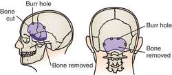
Once symptoms have manifested themselves, the most important thing to determine is whether the shunt is functioning properly and not having intermittent blockages due to a malfunction within the system. In many cases, this can be as easy as measuring the intracranial pressure (ICP) within the skull. Once the functionality of the shunt is assessed, the next step is to consider a volume expansion procedure such as a subtemporal decompression. This procedure involves removing a small section of bone from the skull which allows the ventricle - and the catheter - to expand and relieves the excess pressure.

Saturday, September 9, 2017

What is endoscopic third ventriculostomy (ETV)? (Part 1 of 2)


I. What is an endoscopic third ventriculostomy?

In performing an endoscopic third ventriculostomy or ETV, a neurosurgeon makes a small perforation in the thinned floor of the third ventricle thereby allowing the drainage of cerebrospinal fluid (CSF) out of the blocked ventricular system and into the interpenducular cistern (which is a normal CSF space). This allows CSF within the ventricle in an attempt to bypass an obstruction in the
aqueduct of Sylvius, thereby relieving pressure. The objective of this procedure - technically known as a "Intracranial CSF Diversion" is to normalize pressure on the brain without implanting a shunt. It should be emphasized, however, an ETV is not a cure for hydrocephalus, but, rather, an alternate treatment.

Although open ventriculostomies were performed as early as 1922, they become less common in the
1960's with the advent of shunt systems. Despite the recent improvements in shunting technology and surgical techniques, their use in certain cases remains inadequate. This is due to complications such as blockage, infection, and over-drainage which often require repeated surgeries to permanently correct them. Because of these factors, many neurosurgeons are again recommending an ETV instead of implanting a shunt.

II. New technologies renew interest in ETV

This renewed interest in the use of ETV as an alternative treatment for hydrocephalus is due in a large part to the development of a technology known as neuroendoscopy which allows a neurosurgeon access to areas of the brain inaccessible with traditional surgical techniques. It involves passing a tiny viewing scope into the third ventricle of the brain allowing images of the ventricle to be projected onto a screen located in the operating room.

Typically, the endoscopic catheter is passed through a small hole burred (drilled) in the skull. In some patients (who are already shunted) the neurosurgeon may be able to use the original bone defect made when the shunt was initially placed.

III. Who is a candidate for ETV?

Most physicians seem to agree that there are three (3) factors that lead to a successful ventriculostomy: 1) Patient's age (it is recommended they be over age six (6) years); 2) Prior placement of a shunt; and 3) A diagnosis of non-communicating (obstructed ventricular pathways). Additionally, some doctors have noted a higher success rate in patients with aqueductal stenosis which is the most common cause of congenital hydrocephalus. Aqueductal stenosis, is a result of the long, narrow passageway between the third and fourth ventricles. The end result is an accumulation of fluid upstream from the blockage.

Doctors hypothesize that previous shunt presence possibly allows development of the subarachnoid space and the presence of a functional shunt buys time for the patient while he/she develops absorption abilities. It should be noted, however, that in patients that have been shunted for a number of years, it is often difficult to determine whether or not the hydrocephalus is communicating or non-communicating without undergoing invasive testing.


Additional information: Endoscopic third ventriculostomy

Monday, August 21, 2017

Shunting as a means to manage hydrocephalus


I. Overview

Image result for subarachnoid space
Subarachnoid spaces in the brain

I. Overview

Over the years, the management of hydrocephalus has challenged neurologist, engineers, and medical device manufacturer alike due to the unique nature of cerebrospinal fluid (CSF) dynamics in each person. Known as a "CSF diversion device", a shunt has become the primary therapy used in hydrocephalus management for over sixty (60) years. The shunt, which is surgically implanted within a ventricle in the patient's brain (or in the subarachnoid spaces around the brain), works by diverting the CSF to another part of the body where it is absorbed. This creation of an alternate pathway typically restores the physiological balance between CSF production, flow, and absorption when one or more of these functions has been impaired. Once it has been inserted, valves within the shunt's pathway act like on/off switches, opening when the differential pressure, that is, the pressure difference across the valve, exceeds the valve's opening pressure.
Blog author Walter Little with his
grandparents, James and Sara Sanders,
and Officer Dorsey Goss (1973)

II. What are the complications of shunting?

As I alluded to above, a shunt provides an alternate pathway through which CSF can bypass obstruction(s) in the fluid compartments (ventricles) of the brain. Such a bypass relieves the excess fluid backup the causes hydrocephalus. When both the CSF production and absorption are in balance, the hydrocephalus is considered to be "compensated". In contrast, when production exceeds aborption, complications such as elevated pressure or overdrainage occur and can mimic a malfunctioning shunt.

More detailed information can be found on the Hydrocephalus Association website, but here is a brief overview of some of the most common complications:

Malfunction

As is the case with any piece of equipment, a shunt might break. Additional they are subject to becoming disconnected, migrating (moving), or, most commonly, becoming blocked. (It should be noted that the disconnection and migration are particularly prevalent in children with a shunt due to the child's growth.)

Infection

Shunts can become colonized with bacteria or - in rare instances - fungi which typically occurs at the time the shunt is implanted. In an effort to combat this, some manufacturers have began to add anti-microbial coating to their shunts which appears to be reducing the rate of post-surgical infection.

Material degradation

Originally, barium sulfate (BaSO4) was mixed with silicone to allow the shunt catheter to be visible on an x-ray. These would eventually dissolve making the tubing surface rough. When tissue in-growth occurred to the tubing, it would bind at that location resulting in deterioration and/or breakage. Over time the design of shunt tubing has changed and a clear silicone elastomer now covers the surface of the tubing greatly decreasing the likelihood of degradation.

 Shunt revision(s)

I put an "s" on revision because the average person with hydrocephalus undergoes 2.66 shunt revisions over the course of their lifetime. These can be required at any time to correct one (or more) of the complications outlined above or to compensate for growth in the case of children and young adults. If a blockage is suspected, it must be confirmed by a neurosurgeon who evaluates the implanted system to determine whether the problem is the result of a complete or partial blockage, if a disconnection has occurred, or whether the current system just can't no longer meet the needs of the individual.

Additional information:

Shunt system fact sheet

Thursday, August 17, 2017

What is hydrocephalus? (Part 1 of 2)


Countdown to Washington: 3 weeks and four (4) days

I. What is hydrocephalus?
Taken shortly after my birth,
my hydrocephalus was clearly evident

After completing my latest blog last night, it occurred to me that some might not know and/or understand what hydrocephalus is. It is a condition caused by an abnormal accumulation of cerebrospinal fluid (CSF) in the ventricles (pictured above) of the brain.  The word comes from the Greek hydro (meaning "water") and cephalus (meaning "head). Often, the term "water head" is used in a derogatory sense to make fun of a person suffering from the condition.

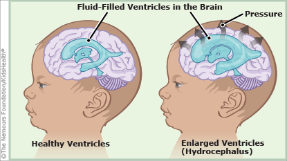
Under normal conditions the CSF - which is produced in the ventricles - circulates through the ventricular system before being absorbed into the blood stream. It is constantly being circulated and serves many functions in ensuring the health of both the brain and the spinal cord. Perhaps one of its most important functions is to surround both the brain and spinal cord and serve as a cushion against injury. In persons with hydrocephalus (or "hydro" for short) there is an imbalance between the amount of CSF produced and the rate at which it is absorbed into the blood stream. As it builds up within the ventricles, it causes them to enlarge and the pressure to increase.

II. What causes hydrocephalus?

First of all, it's important to differentiate that hydrocephalus is a condition and not a disease  (emphasis added) as I have heard people mistakenly say over the years. It can develop for a variety of reasons including as a part of another condition.

Hydrocephalus that is congenital (meaning that it is present at birth as mine was) is thought to be the result of a complex interaction of both genetic and environmental factors. One of the most common causes of hydrocephalus is Aqueductal stenosis where there is a blockage of the cerebral aqueduct known as the Aqueduct of Sylvius.  It is estimated that hydrocephalus occurs in 1.5 per 1,000 births.

In addition to congenital hydrocephalus, it can also be acquired as the result of intraventricular hemorrhage, meningitis, head trauma, a tumor, or a cyst.

Yet a third cause of hydrocephalus - known as normal pressure hydrocephalus or NPH - is prevalent in older adults. Unlike congenital or acquired hydrocephalus, NPH causes little or no appreciable increase in pressure within the ventricles.


For additional information: Hydrocephalus fact sheet