Showing posts with label Ventriculostomy. Show all posts

Monday, September 18, 2017

What is endoscopic third ventriculostomy (ETV)? (Part 2 of 2)

View of the interior of the brain from the right lateral ventricle
In the first part of this blog, I discussed exactly what an endoscopic third ventriculostomy is, what has brought a renewed interest in the procedure, and who makes a good candidate for it. Today I will look at potential complications as well as how success is measured in patients who undergo an ETV.

IV. What are the potential complications of an ETV
Stereotactically guided endoscopy

Thanks to new technology - such as Magnetic Resonance Imaging (MRI), Stereotactically guided endoscopy (pictured at right), flexible fiber optic scopes, and improved tools for manipulation and hemostasis (stoppage of blood flow) the risks associated with an ETV have been minimized. The advent of high-resolution MRI images now allows the neurosurgeon to clearly perceive the absence of CSF through a stenosed or occluded aqueduct, while neuroendoscopic offer unprecedented views from within the ventricular system itself.

The most common complications associated with an ETV are post-procedure fever and bleeding. The fever is the result of a combination of (a) cold light source and monopolar coagulation in the confined volume of the third ventricle which elevates the temperature of the CSF and can cause a fever. Additionally, attempts to perforate the ventricular floor can lead to bleeding, as can damage to the ventricular wall or perforation of the basilar artery. Large bleeds under the third ventricle can be catastrophic, but are rare.

Short-term memory loss is yet another potential complication associated with ETV. This is due to the fact that the procedure can affect both the hypothalamus as well as areas of the mamillary body which is responsible for memory. Over time, however, the individual usually recovers from any short-term memory loss

V. Measuring success of an ETV

"Success" of an ETV is usually considered (by both patients and doctors alike) to be the avoidance of placement of a shunt in a patient who would have otherwise required one. Medical professionals would qualify the procedure as a "success" if their patient exhibits clinical evidence of normal intracranial pressure (ICP) AND structural evidence of stable or decreased ventricular size. (Emphasis added) In the case of a patient who has previously been shunted, the shunted must have either been removed or proved to be non-functional to demonstrate success.

Conversely, most doctors would consider an ETV as a failure if the patient exhibits no change in their clinical symptoms (or ventricular size) or requires the placement of a shunt within days or months of the ETV placement.

Additional information: Endoscopic third ventriculostomy


Saturday, September 9, 2017

What is endoscopic third ventriculostomy (ETV)? (Part 1 of 2)


I. What is an endoscopic third ventriculostomy?

In performing an endoscopic third ventriculostomy or ETV, a neurosurgeon makes a small perforation in the thinned floor of the third ventricle thereby allowing the drainage of cerebrospinal fluid (CSF) out of the blocked ventricular system and into the interpenducular cistern (which is a normal CSF space). This allows CSF within the ventricle in an attempt to bypass an obstruction in the
aqueduct of Sylvius, thereby relieving pressure. The objective of this procedure - technically known as a "Intracranial CSF Diversion" is to normalize pressure on the brain without implanting a shunt. It should be emphasized, however, an ETV is not a cure for hydrocephalus, but, rather, an alternate treatment.

Although open ventriculostomies were performed as early as 1922, they become less common in the
1960's with the advent of shunt systems. Despite the recent improvements in shunting technology and surgical techniques, their use in certain cases remains inadequate. This is due to complications such as blockage, infection, and over-drainage which often require repeated surgeries to permanently correct them. Because of these factors, many neurosurgeons are again recommending an ETV instead of implanting a shunt.

II. New technologies renew interest in ETV

This renewed interest in the use of ETV as an alternative treatment for hydrocephalus is due in a large part to the development of a technology known as neuroendoscopy which allows a neurosurgeon access to areas of the brain inaccessible with traditional surgical techniques. It involves passing a tiny viewing scope into the third ventricle of the brain allowing images of the ventricle to be projected onto a screen located in the operating room.

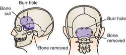
Typically, the endoscopic catheter is passed through a small hole burred (drilled) in the skull. In some patients (who are already shunted) the neurosurgeon may be able to use the original bone defect made when the shunt was initially placed.

III. Who is a candidate for ETV?

Most physicians seem to agree that there are three (3) factors that lead to a successful ventriculostomy: 1) Patient's age (it is recommended they be over age six (6) years); 2) Prior placement of a shunt; and 3) A diagnosis of non-communicating (obstructed ventricular pathways). Additionally, some doctors have noted a higher success rate in patients with aqueductal stenosis which is the most common cause of congenital hydrocephalus. Aqueductal stenosis, is a result of the long, narrow passageway between the third and fourth ventricles. The end result is an accumulation of fluid upstream from the blockage.

Doctors hypothesize that previous shunt presence possibly allows development of the subarachnoid space and the presence of a functional shunt buys time for the patient while he/she develops absorption abilities. It should be noted, however, that in patients that have been shunted for a number of years, it is often difficult to determine whether or not the hydrocephalus is communicating or non-communicating without undergoing invasive testing.


Additional information: Endoscopic third ventriculostomy