Showing posts with label Intracranial. Show all posts

Saturday, September 9, 2017

What is endoscopic third ventriculostomy (ETV)? (Part 1 of 2)


I. What is an endoscopic third ventriculostomy?

In performing an endoscopic third ventriculostomy or ETV, a neurosurgeon makes a small perforation in the thinned floor of the third ventricle thereby allowing the drainage of cerebrospinal fluid (CSF) out of the blocked ventricular system and into the interpenducular cistern (which is a normal CSF space). This allows CSF within the ventricle in an attempt to bypass an obstruction in the
aqueduct of Sylvius, thereby relieving pressure. The objective of this procedure - technically known as a "Intracranial CSF Diversion" is to normalize pressure on the brain without implanting a shunt. It should be emphasized, however, an ETV is not a cure for hydrocephalus, but, rather, an alternate treatment.

Although open ventriculostomies were performed as early as 1922, they become less common in the
1960's with the advent of shunt systems. Despite the recent improvements in shunting technology and surgical techniques, their use in certain cases remains inadequate. This is due to complications such as blockage, infection, and over-drainage which often require repeated surgeries to permanently correct them. Because of these factors, many neurosurgeons are again recommending an ETV instead of implanting a shunt.

II. New technologies renew interest in ETV

This renewed interest in the use of ETV as an alternative treatment for hydrocephalus is due in a large part to the development of a technology known as neuroendoscopy which allows a neurosurgeon access to areas of the brain inaccessible with traditional surgical techniques. It involves passing a tiny viewing scope into the third ventricle of the brain allowing images of the ventricle to be projected onto a screen located in the operating room.
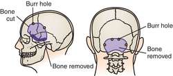
Typically, the endoscopic catheter is passed through a small hole burred (drilled) in the skull. In some patients (who are already shunted) the neurosurgeon may be able to use the original bone defect made when the shunt was initially placed.

III. Who is a candidate for ETV?

Most physicians seem to agree that there are three (3) factors that lead to a successful ventriculostomy: 1) Patient's age (it is recommended they be over age six (6) years); 2) Prior placement of a shunt; and 3) A diagnosis of non-communicating (obstructed ventricular pathways). Additionally, some doctors have noted a higher success rate in patients with aqueductal stenosis which is the most common cause of congenital hydrocephalus. Aqueductal stenosis, is a result of the long, narrow passageway between the third and fourth ventricles. The end result is an accumulation of fluid upstream from the blockage.

Doctors hypothesize that previous shunt presence possibly allows development of the subarachnoid space and the presence of a functional shunt buys time for the patient while he/she develops absorption abilities. It should be noted, however, that in patients that have been shunted for a number of years, it is often difficult to determine whether or not the hydrocephalus is communicating or non-communicating without undergoing invasive testing.


Additional information: Endoscopic third ventriculostomy

Friday, August 25, 2017

Does it hurt? Hydrocephalus and the headaches associated with it (Part 1 of 2)


The other evening I was looking at what I had written about so far and thinking of additional topics when a long-time friend suggested "anything but the typical 'does it hurt' '" that she and I have heard too much as we grew up with a disability and even now as we are in our adult years. Following her comment, I began looking and came up with the idea of doing a blog on hydrocephalus and the associated headaches suffered by some of us.

I. Mechanics of hydrocephalus and headaches

The combination of brain volume (volume for the average adult is 1,260 cubic centimeters), blood volume, and cerebrospinal fluid (CSF) volume determine intracranial pressure or ICP. If one of these increases, pressure in the brain will rise unless one of the others compensates by decreasing. In a person - such as myself - with hydrocephalus this balance is distorted and a unnatural condition takes place. In a person not affected by hydrocephalus, they should have one ounce (1 oz.) of CSF in the ventricles and about four ounces (4 oz.) of CSF surrounding the outside of the brain. When the components of the brain are functioning normally - without a shunt - the brain has the ability to be elastic. This means it is able to compensate accordingly for an increase (or decrease in CSF pressure). In hydrocephalus patient's who have been shunted, this is no longer the case.

No automatic alt text available.
Physiology of hydrocephalus
In studying to present this blog, I learned something that I had never heard before and that is that when our bodies enter rapid eye movement (REM) sleep (typically about 80 - 90 minutes after falling alseep) plateau waves occur resulting in increased intracranial pressure (ICP). In shunted patients who undergo ICP monitoring overnight, those changes are considerably more drastic that with the average person. Those things that would normally be expected to cause an increase in ICP result in huge changes in a shunted patient.

Placement of a shunt results in an unnatural situation. The brain fills the intracranial space while the shunt drains essentially of the available CSF from the ventricles.The end result is a larger-than-normal brain in a fixed (NOT elastic) skull with little room for changes in the ICP (which is characteristic of a person with hydrocephalus. If changes in blood flow occur, resulting in blood volume within the intracranial space, increased intracranial pressure will occur and can result in a headache.

Headaches can also be the result of altered pressures within the skull once a shunt has been implanted. Interestingly, the can occur both if the pressure is too high or too low.

II. Treatment

Like everyone else, both children and adults with hydrocephalus experience periodic headaches. It is the frequency and severity of these that determine whether they are a result of the hydrocephalus. According to Dr. Gordon McComb, Chief of Neurosurgery, Children's Hospital Los Angeles, "If the headaches are getting progressively worse, many times it's an intermittent malfunction of the shunt". Dr. McComb explains that ventricle size is also not always an accurate indication of whether the shunt is working correctly. "The ventricles are going to remain the exact same size whether the pressure is normal or elevated".

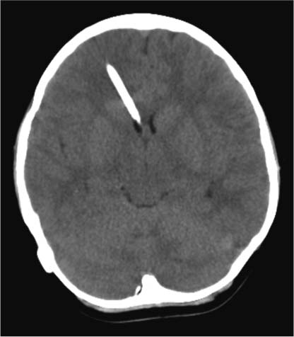
The slit ventricle is the white streak
visible pointing toward the
left side of the brain
With slit ventricle syndrome, small ventricle size is not the cause of the problem. The problem occurs if the shunt becomes clogged and the ventricles don't dilate. If this situation happens repeatedly (accompanied by acceleration in the occurrence of headaches) it can indicate a blockage the builds up and then releases. Replacing the proximal ventricular catheter in the shunt will normally correct this problem.

When hydrocephalus problems experience repeated headaches it is imperative to sketch out a plan of action which involves a conference between the patient and his/her doctor(s). This conference sets up treatment parameters and, if those parameters are exceeded, the medical team moves forward with the replacement of the shunt.


For additional information: Hydrocephalus and headaches